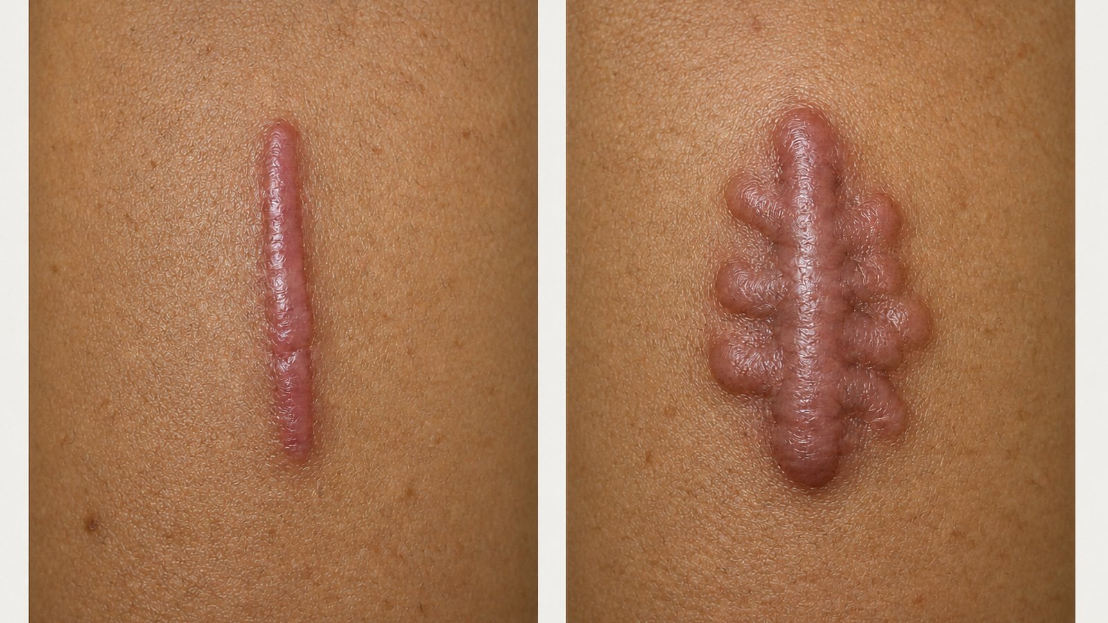
แผลเป็นนูนไม่ได้แปลว่าเป็นคีลอยด์
A raised scar is not automatically a keloid
ทั้งคีลอยด์และแผลเป็นนูนเป็นการสร้างเนื้อเยื่อแผลเป็นมากเกินไป และทั้งคู่ไม่ใช่มะเร็ง ทั้งสองแบบอาจแข็ง นูน แดงหรือคล้ำกว่าผิวรอบข้าง คัน และเจ็บได้เหมือนกัน จึงแยกจากลักษณะภายนอกเพียงอย่างเดียวได้ยาก
Both keloids and hypertrophic scars are benign overgrowths of scar tissue. Both can be firm, raised, red or darker than the surrounding skin, itchy and tender. Appearance alone rarely separates them.
สิ่งที่ช่วยแยกได้ดีที่สุดไม่ใช่ว่าแผลดูรุนแรงแค่ไหน แต่คือแผลลามออกนอกขอบแผลเดิมหรือไม่ และเมื่อเวลาผ่านไปแผลนั้นทำตัวอย่างไร
The most useful distinction is not how dramatic the scar looks, but whether it has crossed the boundary of the original wound, and how it behaves over time.
แผลเป็นนูน — นูนขึ้น
คีลอยด์ — ลามออก
เป็นเพียงวิธีจำง่าย ๆ ไม่ใช่การวินิจฉัยด้วยตัวเอง
Hypertrophic grows up.
Keloid grows out.
A memory aid, not a self-diagnosis.
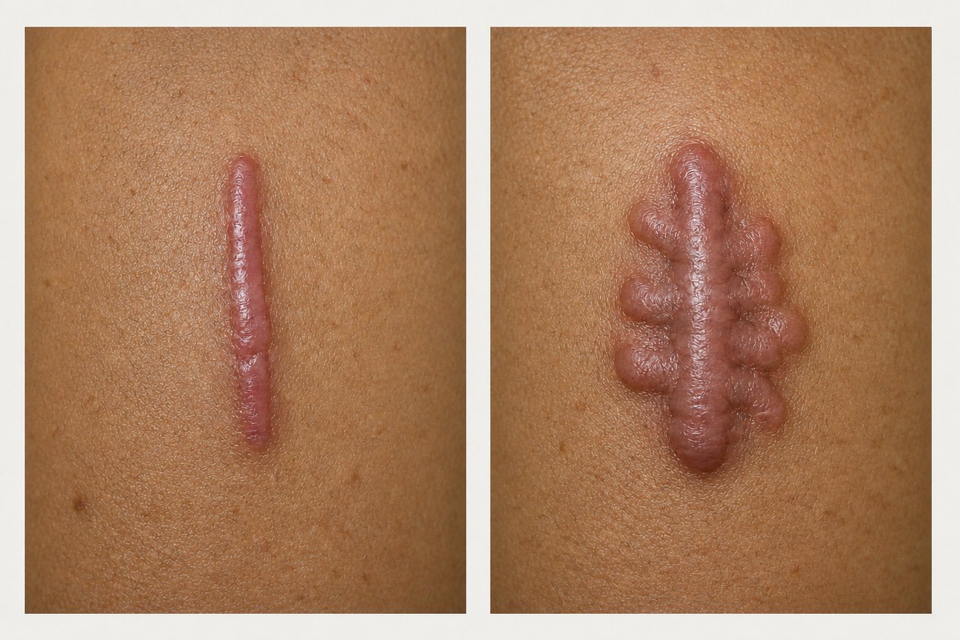
ภาพประกอบเพื่อการศึกษา ไม่ใช่ภาพถ่ายของผู้ป่วยจริง
Educational illustration, not a photograph of a real patient.
ข้อควรระวังคือ แผลเป็นบางรายมีลักษณะก้ำกึ่ง และบางครั้งขอบแผลเดิมมองไม่เห็นแล้ว อีกทั้งยังไม่มีการตรวจเลือดหรือตัวบ่งชี้ใดที่แยกสองภาวะนี้ได้อย่างสมบูรณ์
A caution: some scars sit in between, and the original wound edge is sometimes no longer visible. There is also no blood test or single marker that separates the two perfectly.
ตารางเปรียบเทียบ
Side by side
| จุดสังเกต | แผลเป็นนูน (Hypertrophic) | คีลอยด์ (Keloid) |
|---|---|---|
| การลุกลาม | นูนขึ้น แต่อยู่ในขอบแผลเดิม | ลามออกนอกขอบแผลเดิม เข้าไปในผิวปกติ |
| ช่วงเวลาที่เกิด | มักเริ่มภายใน 4–8 สัปดาห์ | อาจเริ่มหลังจากนั้นหลายเดือนหรือเป็นปี |
| การดำเนินโรค | มักคงที่ และค่อย ๆ ยุบลงได้เอง | มักไม่ยุบเอง และอาจโตต่อเนื่อง |
| บริบทที่พบบ่อย | แผลไฟไหม้ แผลตึง แผลหายช้า มักสำคัญเมื่ออยู่ใกล้ข้อ | หน้าอก หัวไหล่ หลังส่วนบน คอ กราม ติ่งหู |
| การรั้งของแผล | อาจหดรั้งจนขยับข้อได้น้อยลง | ไม่ใช่ลักษณะเด่นของคีลอยด์ |
| Clue | Hypertrophic scar | Keloid |
|---|---|---|
| Growth | Raised, but stays inside the original wound | Extends beyond the wound into normal skin |
| Timing | Usually begins within 4–8 weeks | May begin months or even years later |
| Natural course | Often stabilises and may flatten on its own | Rarely regresses; may keep growing |
| Common context | Burns, high-tension wounds, delayed healing; matters most near joints | Chest, shoulders, upper back, neck or jawline, earlobes |
| Contracture | Can tighten and restrict a joint | Not a classic feature |
ทำไมจึงเกิดขึ้น
Why it happens
ในการหายของแผลตามปกติ ร่างกายจะสร้างคอลลาเจนเพื่อปิดแผล แล้วค่อย ๆ ปรับโครงสร้างคอลลาเจนนั้นเมื่อแผลเป็นเข้าที่ แต่ในทั้งสองภาวะนี้ การอักเสบและการสร้างคอลลาเจนยังคงทำงานมากเกินไป
In normal healing the body lays down collagen to close the wound, then remodels it as the scar matures. In both of these conditions, inflammation and collagen production stay overactive.
ปัจจัยที่มีผล ได้แก่ พันธุกรรม แรงตึงของแผล การอักเสบที่ยืดเยื้อ แผลที่หายช้า และตำแหน่งบนร่างกาย ผู้ที่มีผิวสีเข้มและบางครอบครัวมีความเสี่ยงเป็นคีลอยด์สูงกว่า แต่ทุกคนเป็นได้ทั้งสองแบบ สีผิวเป็นเพียงปัจจัยเสี่ยง ไม่ใช่การวินิจฉัย
Contributing factors include genetics, wound tension, prolonged inflammation, delayed healing and body location. People with darker skin tones, and some families, carry a higher keloid risk, but anyone can develop either type. Skin tone is a risk factor, not a diagnosis.
ไม่ใช่ก้อนนูนทุกก้อนคือแผลเป็น
Not every firm lump is scar tissue
นี่คือประเด็นด้านความปลอดภัยที่สำคัญที่สุดของบทความนี้ ก้อนเนื้อบางชนิดและมะเร็งผิวหนังบางชนิดที่พบไม่บ่อย เช่น dermatofibrosarcoma protuberans (DFSP) มะเร็งเซลล์สความัส และเมลาโนมาชนิดไม่มีสี สามารถมีหน้าตาคล้ายคีลอยด์ได้
This is the most important safety point in this article. Some benign growths and uncommon skin cancers, including dermatofibrosarcoma protuberans (DFSP), squamous cell carcinoma and amelanotic melanoma, can look like a keloid.
มีรายงานผู้ป่วยจำนวนหนึ่งที่ DFSP ถูกเข้าใจผิดว่าเป็นคีลอยด์และถูกผ่าตัดออกในฐานะแผลเป็น ทำให้การวินิจฉัยล่าช้า หากประวัติหรือลักษณะของก้อนไม่เข้ากับแผลเป็นทั่วไป แพทย์อาจแนะนำให้ตัดชิ้นเนื้อส่งตรวจก่อนเริ่มรักษาแผลเป็น
There are documented cases of DFSP being mistaken for a keloid and excised as a scar, delaying the real diagnosis. If the history or appearance does not fit an ordinary scar, a doctor may recommend a biopsy before any scar treatment begins.
ควรพบแพทย์เพื่อประเมิน แทนการรักษาเอง หากมีข้อใดข้อหนึ่งต่อไปนี้
ไม่มีประวัติบาดแผล การเจาะ การผ่าตัด สิว หรือการอักเสบที่ตำแหน่งนั้นมาก่อน · ก้อนโตเร็วผิดปกติ หรือหน้าตาไม่เหมือนแผลเป็นอื่นบนตัวเรา · การวินิจฉัยยังไม่ชัดเจน หรือเคยรักษาแบบ “แผลเป็น” แล้วไม่ได้ผลซ้ำ ๆ · มีอาการปวด คัน ผิวแตก หรือขยับได้น้อยลงจนกระทบชีวิตประจำวัน
Have it assessed rather than self-treating if any of these apply
There was no injury, piercing, operation, acne lesion or inflammation at that site · the lump is growing unusually fast, or looks unlike your other scars · the diagnosis is uncertain, or previous "scar" treatment has repeatedly failed · pain, itch, skin breakdown or restricted movement is affecting daily life.
แนวทางการรักษาคือแผน ไม่ใช่ผลิตภัณฑ์ชิ้นเดียว
Treatment is a plan, not a single product
เป้าหมายที่เป็นจริงคือการทำให้ดีขึ้นอย่างมีความหมาย ได้แก่ คันและเจ็บน้อยลง แผลแบนและนุ่มขึ้น ขยับได้ดีขึ้น และลดโอกาสกลับมาเป็นซ้ำ ปัจจุบันยังไม่มีวิธีใดรับประกันว่าแผลเป็นจะหายไปทั้งหมด
The realistic goal is meaningful improvement: less itch and pain, a flatter and softer scar, better movement, and a lower chance of recurrence. No current method can promise a scar will disappear entirely.
- 1. ดูแลแผลให้หายดีก่อน — ลดแรงตึงที่หลีกเลี่ยงได้ และดูแลให้แผลหายโดยไม่มีภาวะแทรกซ้อน เริ่มใช้ผลิตภัณฑ์ดูแลแผลเป็นเมื่อผิวปิดสนิทแล้วเท่านั้น
- 2. ทำให้แผลสงบและแบนลง — แผ่นหรือเจลซิลิโคน และการกดทับ เป็นทางเลือกที่ใช้กันในแผลเป็นบางลักษณะ ซิลิโคนเป็นทางเลือกที่สมเหตุสมผล แต่ควรทราบว่างานวิจัยคุณภาพสูงยังมีจำกัด
- 3. รักษาเนื้อเยื่อแผลเป็นที่ยังทำงานอยู่ — แพทย์อาจใช้การฉีดยาสเตียรอยด์ การจี้เย็น ยา 5-fluorouracil หรือเลเซอร์บางชนิด มักใช้ร่วมกันและต้องทำหลายครั้ง
- 4. วางแผนการผ่าตัดอย่างรอบคอบ — การผ่าตัดแก้ไขช่วยได้ในแผลเป็นนูนบางราย แต่สำหรับคีลอยด์ ต้องวางแผนป้องกันการกลับเป็นซ้ำตั้งแต่ก่อนผ่าตัด การผ่าตัดอย่างเดียวมักไม่ใช่กลยุทธ์ที่ดี
- 1. Protect the healing wound — reduce avoidable tension and support uncomplicated healing. Start scar products only once the skin has fully closed.
- 2. Calm and flatten — silicone gel or sheeting and pressure are common options for selected scars. Silicone is a reasonable choice, though high-quality trial evidence remains limited.
- 3. Treat active scar tissue — corticosteroid injection, cryotherapy, 5-fluorouracil or selected lasers, often in combination and across several sessions.
- 4. Plan surgery carefully — revision helps selected hypertrophic scars. For a keloid, the recurrence-prevention plan must be decided before excision; surgery alone is usually a poor strategy.
ทำไมคีลอยด์จึงกลับมาเป็นซ้ำ
Why keloids come back
การตัดคีลอยด์ออกคือการสร้างแผลใหม่ ซึ่งเป็นสัญญาณเดียวกับที่กระตุ้นให้เกิดแผลเป็นผิดปกติตั้งแต่แรก ด้วยเหตุนี้ การผ่าตัดและแผนป้องกันการกลับเป็นซ้ำจึงต้องมาคู่กันเสมอ
Excising a keloid creates a fresh wound, which is exactly the signal that triggered the abnormal scarring in the first place. That is why the operation and the recurrence-prevention plan belong together.
45–100%
ช่วงอัตราการกลับเป็นซ้ำที่มีรายงาน เมื่อผ่าตัดคีลอยด์ออกเพียงอย่างเดียว ตัวเลขนี้มาจากการสรุปในแนวทางเวชปฏิบัติ ไม่ใช่การทำนายรายบุคคล ความเสี่ยงเปลี่ยนไปตามตำแหน่งบนร่างกาย ลักษณะของแผลเป็น แรงตึงจากการผ่าตัด ระยะเวลาติดตาม และวิธีรักษาที่เสริมหลังผ่าตัด
45–100%
The reported range of recurrence after keloid excision alone. This figure comes from guideline summaries and is not a personal prediction. Risk shifts with body site, scar biology, surgical tension, length of follow-up, and what treatment is added afterwards.
การฉีดสเตียรอยด์หลังผ่าตัด การกดทับหรือซิลิโคนในตำแหน่งที่เหมาะสม และการฉายรังสีในคีลอยด์ที่คัดเลือกมาแล้วว่ายากต่อการรักษา สามารถช่วยลดการกลับเป็นซ้ำได้ แต่ไม่มีวิธีใดทำให้เป็นศูนย์ การฉายรังสีหลังผ่าตัดไม่ใช่การรักษาลำดับแรกตามปกติ และต้องพูดคุยถึงข้อดีข้อเสียเป็นรายบุคคลในสถานพยาบาลเฉพาะทาง
Postoperative corticosteroid, pressure or silicone in suitable locations, and radiotherapy for carefully selected difficult keloids can all reduce recurrence, though none reduces it to zero. Postoperative radiotherapy is not routine first-line care and requires an individual discussion of benefits and risks in a specialist setting.
แผลเป็นนูนมักมีการดำเนินโรคที่ดีกว่า แต่หากยังมีแรงตึงต่อเนื่องหรือมีการหดรั้งจนขยับได้น้อยลง ก็ยังจำเป็นต้องรักษา
Hypertrophic scars generally follow a more favourable course, though persistent tension or contracture may still need treatment.
สรุป
The bottom line
ถ้าแผลเป็นนูนยังอยู่ในขอบแผลเดิมและค่อย ๆ สงบลง แสดงว่ามีพฤติกรรมแบบแผลเป็นนูน ถ้าลามออกนอกขอบแผลเดิมและยังโตต่อเนื่อง แสดงว่ามีพฤติกรรมแบบคีลอยด์
If a raised scar stays inside the original injury and gradually settles, it is behaving like a hypertrophic scar. If it crosses the old boundary and keeps growing, it is behaving like a keloid.
แต่เมื่อขอบแผลไม่ชัดเจน การเติบโตผิดปกติ หรือกำลังพิจารณาจะรักษา ขั้นตอนที่ปลอดภัยที่สุดคือการได้รับการวินิจฉัยที่ถูกต้อง ไม่ใช่การลองผลิตภัณฑ์ชิ้นถัดไป
But when the boundary is unclear, the growth is unusual, or treatment is being considered, the safest next step is a proper diagnosis, not another over-the-counter promise.
ปรึกษาแพทย์เฉพาะทางConsult a specialist
ทุกแผนการรักษาเริ่มต้นจากการตรวจประเมินโดยศัลยแพทย์ตกแต่ง เพื่อออกแบบวิธีที่เหมาะกับลักษณะเฉพาะของแต่ละคน
Every treatment plan begins with an assessment by a plastic surgeon, so the approach fits your individual anatomy rather than a template.
นัดหมายผ่าน LINE →Book via LINE →แหล่งอ้างอิงReferences›
- Ogawa R, et al. Diagnosis and Treatment of Keloids and Hypertrophic Scars — Japan Scar Workshop Consensus Document 2018. Burns & Trauma. 2019;7:39. doi:10.1186/s41038-019-0175-y
- Limandjaja GC, et al. Hypertrophic scars and keloids: overview of the evidence and practical guide for differentiating between these abnormal scars. Experimental Dermatology. 2021;30:146–161. doi:10.1111/exd.14121
- Nast A, et al. S2k guidelines for the therapy of pathological scars — Update 2020. J Dtsch Dermatol Ges. 2021;19:312–327. doi:10.1111/ddg.14279
- Gold MH, et al. Updated international clinical recommendations on scar management: part 2. Dermatologic Surgery. 2014;40:825–831.
- O'Brien L, Jones DJ. Silicone gel sheeting for preventing and treating hypertrophic and keloid scars. Cochrane Database of Systematic Reviews. 2013;(9):CD003826. doi:10.1002/14651858.CD003826.pub3
- Tian F, et al. Silicone gel sheeting for treating keloid scars. Cochrane Database of Systematic Reviews. 2023;(1):CD013878. doi:10.1002/14651858.CD013878.pub2
- Keloid and Hypertrophic Scar — Treatment & Management. Medscape / StatPearls (recurrence after excision alone reported at 45–100%). https://emedicine.medscape.com/article/1057599-treatment
- Case reports of dermatofibrosarcoma protuberans initially treated as keloid. National Library of Medicine (PMC). https://pmc.ncbi.nlm.nih.gov/articles/PMC5789558/
บทความนี้จัดทำขึ้นเพื่อให้ความรู้ทั่วไป ไม่ใช่การวินิจฉัยหรือคำแนะนำการรักษาเฉพาะบุคคล ผลลัพธ์ของแต่ละบุคคลแตกต่างกันตามลักษณะทางกายวิภาค การตรวจประเมินโดยแพทย์จึงจำเป็นเสมอ หากมีอาการผิดปกติ ควรปรึกษาแพทย์โดยตรง
This article is general education, not a diagnosis or personal treatment advice. Individual results vary with anatomy, so an in-person assessment by a doctor is always necessary. If you notice anything unusual, please consult a doctor directly.
← กลับไปหน้าข่าวสารและบทความ← Back to news and articles